


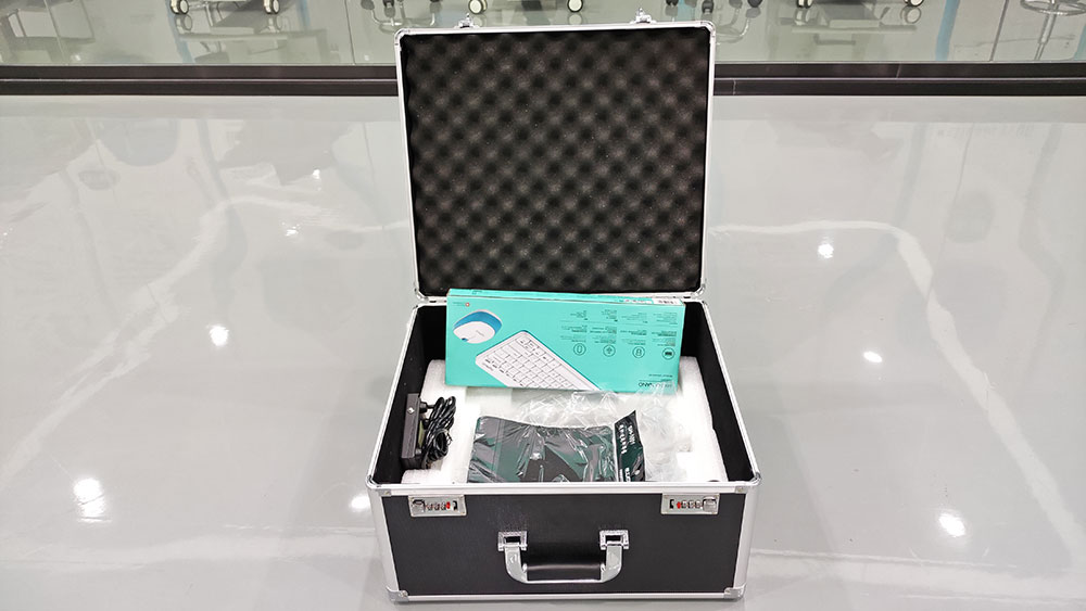
Feature:
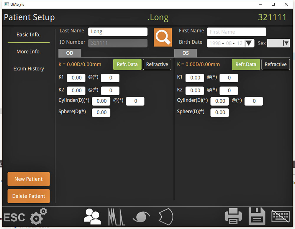
Retiwave-1000 is digital ultrasonic system for ophthalmology. With advanced intelligent digital software the parameters of freezed and stored images could be adjusted voluntarily.Scan A measures data for every part of the eyeball as anterior chamber depth, lens thickness,axial length and so on which are needed in ophthalmic surgeries, and calculates IOL by axial length. Scan B displays profile images of the eyeball clearly and directly. Scanning anatomical formsand nidi inside the eyeball, doctors can diagnose accurately for examination of cataract, vitreousbody disease, ocular trauma, detachment of retina or choroid, macula disease, and intraoculartumor, etc..
 |
 |
 |
 |
 |
 |
| Technical Data | |
| A Probe frequency | 10MHz |
| A Probe Measuring model | Contact/immersion(optional) |
| A Probe Measuring range | Over 5mm~40mm |
| A Probe Measuring error | ≤0.05mm |
| A Probe Measuring type | Length of optic axis, automatic calculation, result analysis |
| A Probe Image freezing | Image freezing auto, auto+ preservable, manual |
| Intraocular lens formula | SRK-T, SRK II, HOLLADAY,BINKHORST-IIHOFFER-Q, HAIGISI |
| Gain | 20~110dB |
| Time gain adjustment | 0~30dB |
| B Probe Frequency | 10Mhz |
| B Probing Depth | ≥50mm |
| Axial Resolution | ≤ 0.2mm |
| Lateral Resolution | ≤0.3mm |
| Gain Adjustment | 20-110db |
| Time Gain Adjustment | 0-30db |
| Operation Mode | Eyecup, gel, aqueous capsule |
| Post Processing Measuring Tools | Area, Length, Mark and Annotation |
| Reviews | 100 images available |
Send Inquiry
Please fill out the form below, and write down your requirements. The field marked with red * is required.


