Feature:
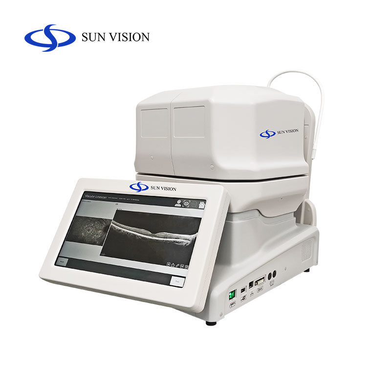
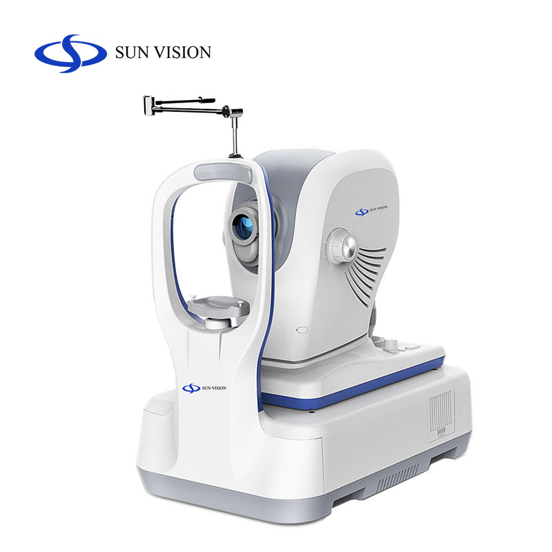
SV-800 is a new generation of HD SD-OCT, with upgraded function, clear image and smooth system. It is equipped with professional analysis software to accurately identify retinal diseases, help screen and reduce the missed diagnosis of the initial examination, which can greatly improve the clinical use efficiency.
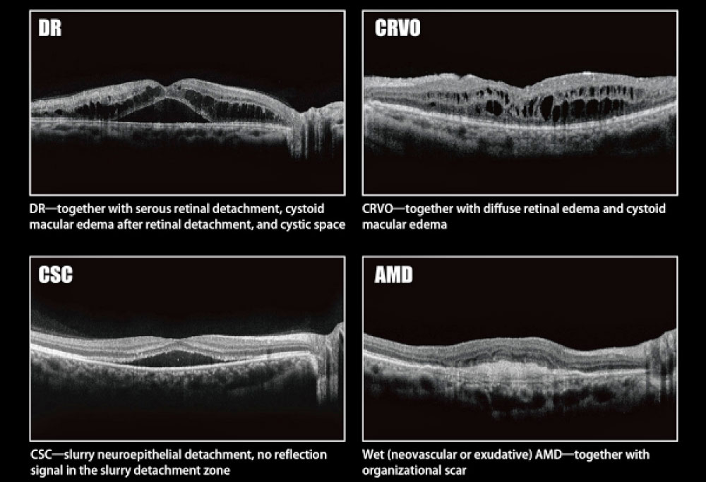
It uses LSLO technology, with up to 2.65mm scan depth, and the lateral resolution of retinal fundus image is up to 5μm. Equipped with professional analysis software, it can obviously show the macular thickness under the macular thickness analysis model, which helps accurately identify clinical macular diseases.
Wide to the Edge of Vision
Large scanning range, clear macular area and optic disc area at a glance.
Auto Focus
SV-800 can automatically complete the tracking of fundus and macula, detect and calibrate the central part of the pupil, detect and adjust the focus and fault position, and display retina layers of high-definition. The whole acquiring time is limited to 5 seconds, greatly saving the diagnosis time.
Scan Mode
SV-800 has a variety of scan modes, including area scan, HD one line scan and multilines scan.
Area scan: 512*64, range 6mm*6mm, range 12mm*12mm
HD one line scan: 2048*30, length 6mm; 1024*30, length 12mm
Multilines scan: 5 lines parallel scan, Radiation scan, Circular scan


| Technical Data | |
| Scan light source | |
| Central wavelength | 843nm |
| Scanning speed | 20KHz |
| Maximum scanning depth | 2.65mm |
| Scanning range | 13mm×13mm |
| Light power | ≤750μw (at the cornea) |
| Refractive compensation range | -20D~+20D |
| Resolution(Optic) | |
| Axial resolution | ≤5μm |
| Horizontal resolution | ≤13μm |
| Fundus image | |
| Linear scanning ophthalmoscope (LSO) | |
| Central wavelength | 780nm |
| Maximum imaging range | ≥45° |
| Power | ≤1.5mW |
| Scan mode | |
| Cube scan | 6mm×6mm,12mm×12mm |
| Multi-line scan | five-line scan, radial scan, scan length 6mm/12mm |
| High list line scan | 6mm/12mm |
| Ring scan | diameter 3mm |
| Analysis functions | Macular thickness analysis, optic disc RNFL thickness analysis, follow-up analysis |
| Anterior segment analysis: corneal thickness and atrial Angle were measured | |
| PC | |
| Hard disc | 2T |
| CPU | I7-8700 |
| GPU | RTX2060 6G |
| RAM | DDR4 16G |
| Display | 24-inch LCD screen |